[SLIPPAGE] no. 1 | spring 2013 | THE CONFLUENCE OF SCIENCE AND ART
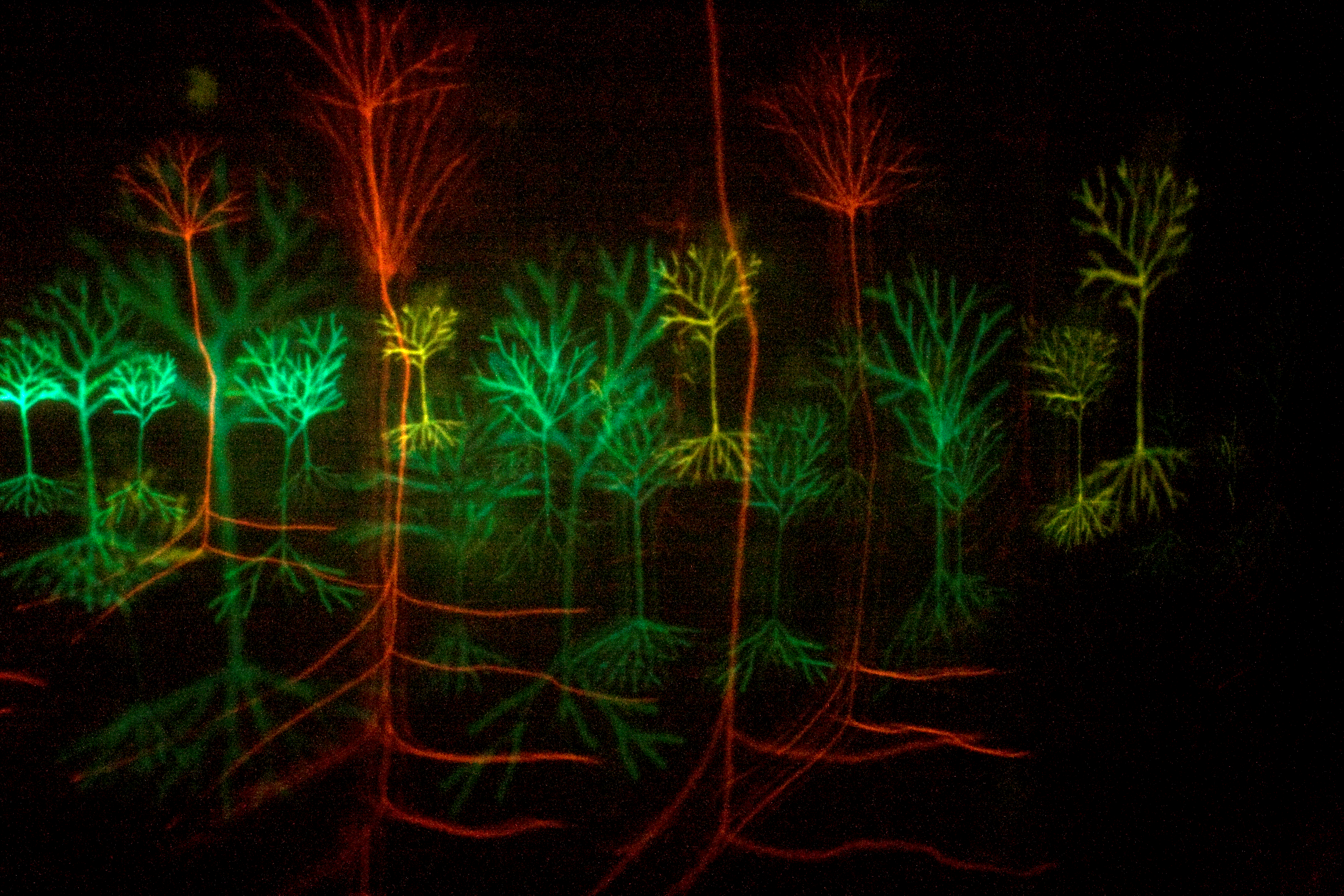
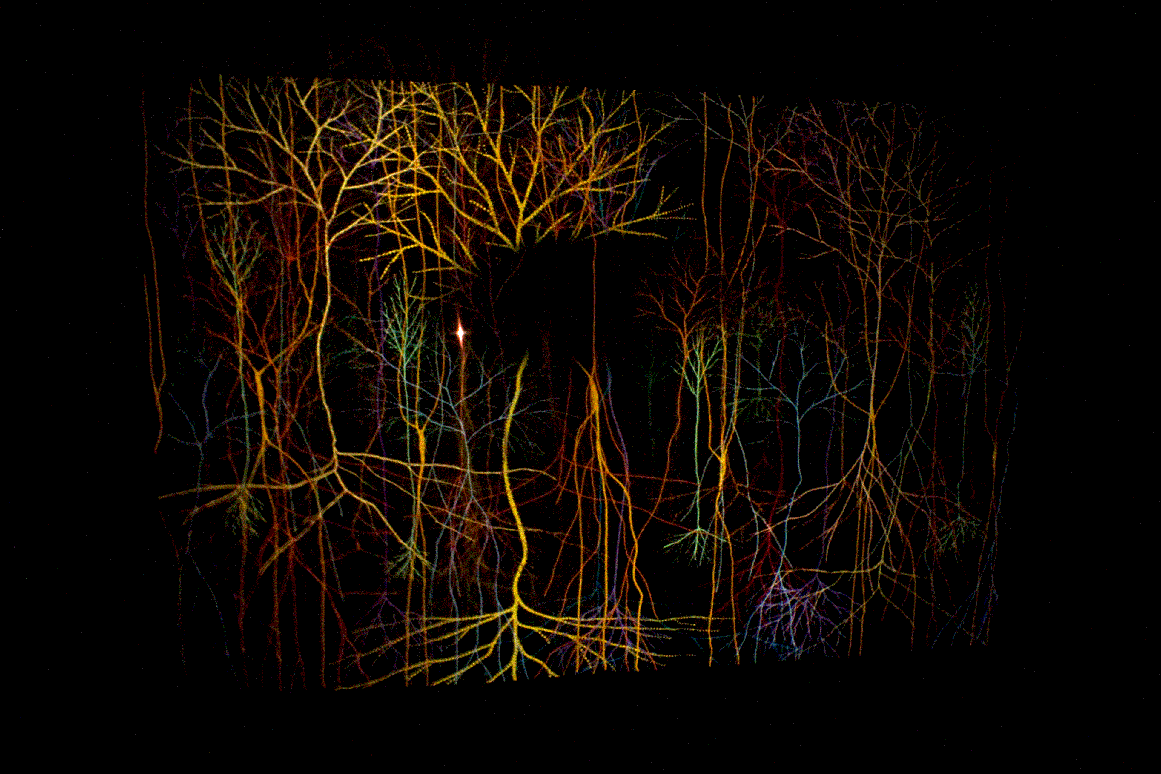
Magic Forest series, by Andrew Carnie
What inspired the work was the ever-growing awareness that the brain is not a static organ. In my youth, I was always told that the brain was ‘fixed’ after the age of 18 or 19 years of age, but current research I indicates otherwise. I was particularly impressed by Richard Wingate’s work on the developing brains of chicks. It was my ongoing conversations with Richard that really made the development of the Magic Forest series possible. What I saw in his QuickTime movies was the structuring of the brain and its neurons over time, an amazing flow of neurons into their working places in the brain. It was incredible to think that despite the time period from the chick's conception to its hatching being only 21 or 22 days, within a few minutes after a chick has pecked its way out of its egg, it can already stand, see, and move around. All of the neurons that make this possible have been created and guided into place over this incredibly short period of time!
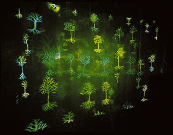
Magic Forest, 2002, is a slowly developing slide dissolve work. The piece takes about 25 minutes to unravel its developing storyline. Two projectors, which sit at opposite ends of a darkened exhibition space at least 6 meters by 4 meters in size, project a sequence of images onto three semi-translucent screens that lie between them. The projected slides rise from one side and then dissolve into images rising from the other side. The final work is a dream-like journey through a forest of developing, ever-expanding neurons.

I was also influenced by the work of Prof Eleanor Maguire at Wellcome Department of Imaging Neuroscience, Institute of Neurology, University College, London. Through fMRI brain scans of London taxi drivers, she was discovering the growth and development of the hippocampus late in life as these taxi drivers gained the "Knowledge", the "Knowledge" being an unbelievably thorough acquaintance of London streets that every black cab driver has to know. The hippocampus is the brain's centre for mapping and spatial awareness. In comparative studies, bus drivers who only have to learn a couple of routes do not have such a large expansion of the hippocampal region.

So Magic Forest was created as a response to this amazing development that also goes on in us; the proliferation of neuron cells, the movement of them, the refinement and organization of the cells within the brain and its continuation over our life time. The work references all these changes as the brain develops, as the projected images slowly sweep from one side to the other on the voile screens. In a way, the screens correspond to the layers of the brain as seen down the sophisticated imaging system that let us see this world. It is fitting that the screens move as viewers pass by, giving life to the work. The end of the work is envisaged as a collapse of the brain to allow the cycle of life to restart through a stroke or some other trauma to the brain.

Richard Wingate uses a confocal microscope to see the neuron cells stained with GFP’s, Green Fluorescent Proteins, within the sections of brain. The sections are kept alive and growing in vitro for some 24 hours. As the cells develop, Richard takes sets of sequential photos of the pattern of neuron development in the chick's brain. These photos are then stacked and compiled into QuickTime movies; when projected, these movies show how the neurons are moving into position in the brain. Hundreds of glowing neurons are moving through the brain's matter, like in a fast-forwarded film of car headlights streaming through a city at night.

Richard switches the particular proteins that attract the growing neurons on and off at different points in the developmental cycle to find out what controls the movement of these neurons. The movies help prove what effect switching on and off any protein has. When particular proteins are switched off at particular times the cells are seen to get lost! They do not know where they are supposed to go in the developing brain; the wiring becomes hectic. Eventual comprehension of the how the protein switches work may lead to cures for some debilitating human disorders.
Richard’s QuickTime videos of chick brains growing in vitro show individual neurons lit by Green Fluorescent Proteins moving to their destinations in the brain. They are an extraordinary glimpse into the ever-changing brain.
I also looked at the work of several early anatomists, particularly the sketches of Spanish anatomist Santiago Ramon Y Cahal, whose drawings of the structure of the brain are particularly exquisite, and the diagrams of Camilio Golgi, the Italian physician and pathologist who first identified the brain as containing neurons using the silver nitrate stain.

Magic Forest was made for the exhibition Head On in 2002, at the Science Museum in London, England. Since then it has been shown at the Rotterdam Film Festival, Holland, 2003; the show Mensbeeld at the Natuurmuseum, Rotterdam in 2004; in the Simply Complex exhibit at the Design Museum in Zurich in 2005; in the Neuroculture exhibit at the Westport Art Centre, Connecticut, USA in 2006; in Exit Art, New York in 2008; in the fabulous exhibition Images of the Mind exhibition at the Dresden Hygiene Museum; and the Morevska Gallery in Brono. Two articles that refer to Magic Forest have appeared in the prominent science journal Nature. This work was produced in collaboration with Wellcome Trust, London, and Arts and Humanities Research Council. (A static version of the time-based piece is now based at the Wellcome Trust Headquarters, London.)
Images and text courtesy of the artist and GV Art Gallery, London